Linking Chromatin Organization and Congenital Heart Disease
A study links TBX5 haploinsufficiency and altered chromatin organization in iPSC-derived cardiomyocytes at single-cell resolution to congenital heart disease.
Linking Altered Chromatin Organization to Mutations in TBX5 and Disease Development
Haploinsufficient mutations in transcription factor genes reduce gene dosage and induce human developmental disorders that affect diverse tissues and cell types by disrupting transcriptional regulation and subsequent lineage specification. Although lineage-constrained transcription factors can directly regulate 3D chromatin organization during cellular differentiation, we still know little regarding how transcription factor haploinsufficiency affects cell-type-specific gene regulation during this time.
Previous studies from researchers led by Benoit G. Bruneau defined the gene expression profile alterations induced by reduced lineage-constrained transcription factor dosage in atrial and ventricular cardiomyocytes differentiated from wild-type induced pluripotent stem cells (iPSCs) and those containing heterozygous loss-of-function or homozygous loss-of-function mutations in the TBX5 gene (Kathiriya et al., 2021 and Kathiriya et al., 2026). Haploinsufficient mutations in the gene encoding the T-box transcription factor TBX5 - a master regulator of cardiac gene expression required for normal heart patterning and morphogenesis (Bruneau et al.) – cause the majority of penetrant congenital heart disease in humans (Basson et al. and Li et al.). Subsequent TBX5-focused research led by Katherine S. Pollard and Benoit G. Bruneau aimed to explore whether alterations in 3D chromatin organization induced the observed transcriptional dysregulation and how TBX5 influences chromatin in a lineage-restricted manner during the differentiation of human iPSCs into cardiomyocytes. The findings of this research, published recently in Science (Grant and Kuang et al.), now reveal that cell type-specific 3D chromatin organization displays sensitivity to lineage-restricted transcription factor dosage and suggest a mechanism by which reduced transcription factor dosage contributes to congenital heart disease development.
Paired-Tag technology from Epigenome Technologies generates joint epigenetic and transcriptomic profiles at single-cell resolution and detects histone modifications and RNA transcripts in nuclei with efficiency comparable to single-nucleus RNA-seq/ChIP-seq assays. Could an in-depth analysis of histone modification and transcriptomic profiles from the same single cells during cardiomyocytic differentiation of iPSCs, enabled by Paired-Tag integration, provide deeper insight into how TBX5 loss affects the chromatin environment and gene expression and potentially promotes the onset of congenital heart disease?
TBX5 Haploinsufficiency Induces Gene Expression Alterations via 3D Chromatin Reorganization
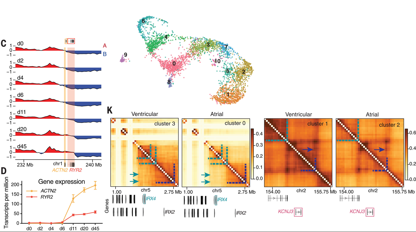
Initial analysis of wild-type human iPSCs during their differentiation into atrial and ventricular cardiomyocytes revealed extensive, dynamic chromatin reorganization; however, the bulk and single-cell techniques employed in this comprehensive study also revealed significant differences between cardiomyocyte types. Specific analysis of TBX5 revealed enrichment at chromatin structures (e.g., topologically associating domain boundaries and chromatin loop anchors) during cardiomyocyte differentiation of iPSCs; however, subsequent analysis of iPSCs with one copy or no copies of TBX5 revealed interesting differences in chromatin organization. The authors observed significant alterations in the higher-order organization of chromosomal compartments, topologically associating domains, and chromatin loops upon the loss of one TBX5 allele that become further exacerbated upon complete TBX5 loss. Notably, in-depth single-cell analyses also revealed varied responses across individual cells.
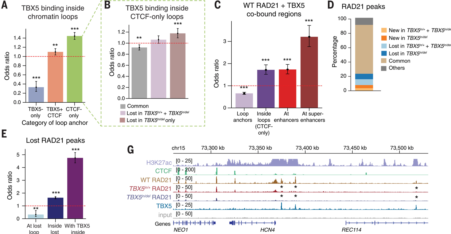
The study also helped to identify regions of the genome sensitive to TBX5 dosage, with changes in TBX5-dependent gene expression strongly associated with alterations in chromatin loops. Finally, while the authors noted that TBX5 loss did not significantly affect CTCF (CCCTC-binding factor; known to support chromatin looping but inhibit cohesin-dependent loop extrusion), they found reduced cohesin occupancy at TBX5-bound enhancers in chromatin loops, which blocked chromatin remodeling during iPSC differentiation into cardiomyocytes. Overall, these fascinating data now provide a potential mechanism for disrupted loop formation in response to TBX5 loss.
Haploinsufficient Transcription Factors, Chromatin Reorganization, and Disease Development: The Next Steps
Overall, this fascinating study reveals how haploinsufficiency in a lineage-restricted transcription factor such as TBX5 can prompt dysregulated gene expression and support the development of congenital heart disease. While mechanisms of haploinsufficiency remained poorly understood, changes in chromatin accessibility had been posited as a possible mechanism (Kathiriya et al., 2026 and Naqvi et al.); here, the authors highlight the implication of 3D chromatin reorganization, which may reflect a generalizable mechanism underpinning the developmental processes dysregulated in human syndromes (Seidman & Seidman). The authors note that we now need additional research to explore this hypothesis using additional haploinsufficient transcription factors and developmental disorders affecting additional cell lineages.
The implementation of Paired-Tag technology from Epigenome Technologies, which generates joint epigenetic and transcriptomic profiles at single-cell resolution and detects histone modifications and RNA transcripts in individual nuclei with efficiency comparable to single-nucleus RNA-seq/ChIP-seq assays, has the potential to provide deeper insight into such research aims. Could the simultaneous single-cell analysis of histone modifications and transcriptomic profiles have contributed to deepening our understanding of how lineage-specific chromatin organization contributes to haploinsufficient transcription factor-mediated transcriptional dysregulation, failed cellular differentiation, and congenital heart disease development?
By Stuart P. Atkinson
Page Title
Meta Summary
Tag
SEO: Chromatin Organization (Add "Chromatin Organization" to the image name and image Alt tag)